Physiotherapy in Toronto for Pediatric Issues
Welcome to In Balance Physiotherapy’s Guide to Perthes Disease of the Hip.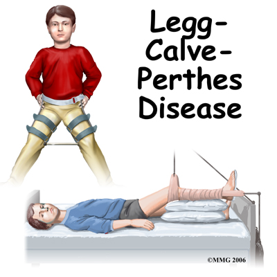 Perthes disease is a condition that affects the hip in children between the ages of four and eight. The condition is also referred to as Legg-Calve-Perthes disease in honor of the three physicians who each separately described the disease. In this condition, the blood supply to the growth center of the hip (the capital femoral epiphysis) is disturbed, causing the bone in this area to die. The blood supply eventually returns, and the bone heals. How the bone heals determines what problems the condition will cause in later life. Perthes disease may affect both hips. In fact, 10 to 12 percent of the time the condition is bilateral (meaning that it affects both hips). This condition can lead to serious problems in the hip joint later in life.
This guide will help you understand:
Perthes disease is a condition that affects the hip in children between the ages of four and eight. The condition is also referred to as Legg-Calve-Perthes disease in honor of the three physicians who each separately described the disease. In this condition, the blood supply to the growth center of the hip (the capital femoral epiphysis) is disturbed, causing the bone in this area to die. The blood supply eventually returns, and the bone heals. How the bone heals determines what problems the condition will cause in later life. Perthes disease may affect both hips. In fact, 10 to 12 percent of the time the condition is bilateral (meaning that it affects both hips). This condition can lead to serious problems in the hip joint later in life.
This guide will help you understand:
- what part of the hip is involved
- what causes the condition
- what treatment options are available
- what In Balance Physiotherapy’s approach to rehabilitation is
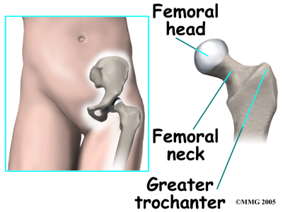
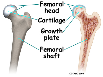
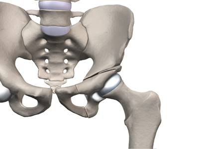
 In the growing child, there are special structures at the end of most bones called growth plates. The growth plate is sandwiched between two special areas of the bone called the epiphysis and the metaphysis. The growth plate is made of a special type of cartilage that builds bone on top of the end of the metaphysis and lengthens the bone as we grow. In the hip joint, the femoral head is one of the epiphyses of the femur.
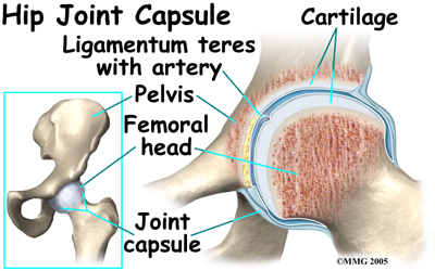
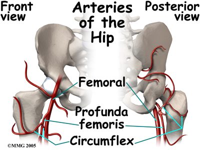
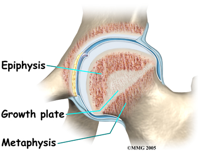
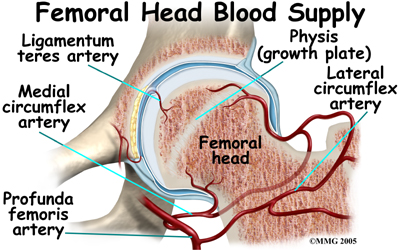 The capital femoral epiphysis is somewhat unique. It is one of the few epiphyses in the body that is inside the joint capsule. (The joint capsule is the tissue that surrounds the joint.) The blood vessels that go to the epiphysis run along the side of the femoral neck and are in danger of being torn or pinched off if something happens to the growth plate. This can result in a loss of the blood supply to the epiphysis
 Children who have abnormal blood clotting (a condition called thrombophilia) may also have a higher risk of developing Perthes disease. These children have blood that clots easier and quicker than normal. This may lead to blood clotting that blocks the small arteries going to the femoral head. As a result of new evidence, the certainty of thrombophilia as a cause of Perthes is now under debate. This will remain an area of study until scientists clear up the significance of thrombophilia as a possible cause of Perthes.
There is some new evidence that Perthes disease may be genetic as a result of a mutation (abnormal change) in the type II collagen (fibers that make up soft tissue structures). Previously there was no known increase in risk for children whose parent had Perthes disease as a child, but this belief may no longer be accurate.
Studies among Asian families who have many family members with this disease have been found with this mutation in the type II collagen gene. Scientists think that the mutation results in weakening of the hip joint cartilage that also affects the blood vessels within the cartilage.
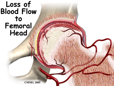
Whatever the true cause of ischemia (lack of blood to the area), the result is bone death (called necrosis) of the femoral head. Without a normal blood supply, the bone loses its strength and shape. The loss of bone density and softening of the head result in a femoral head that is misshaped. With the hip supporting the weight of the body, tiny microfractures in the soft, necrotic bone fail to heal. This is another reason why normal wear and tear results in a deformity.
Children who have abnormal blood clotting (a condition called thrombophilia) may also have a higher risk of developing Perthes disease. These children have blood that clots easier and quicker than normal. This may lead to blood clotting that blocks the small arteries going to the femoral head. As a result of new evidence, the certainty of thrombophilia as a cause of Perthes is now under debate. This will remain an area of study until scientists clear up the significance of thrombophilia as a possible cause of Perthes.
There is some new evidence that Perthes disease may be genetic as a result of a mutation (abnormal change) in the type II collagen (fibers that make up soft tissue structures). Previously there was no known increase in risk for children whose parent had Perthes disease as a child, but this belief may no longer be accurate.
Studies among Asian families who have many family members with this disease have been found with this mutation in the type II collagen gene. Scientists think that the mutation results in weakening of the hip joint cartilage that also affects the blood vessels within the cartilage.
Whatever the true cause of ischemia (lack of blood to the area), the result is bone death (called necrosis) of the femoral head. Without a normal blood supply, the bone loses its strength and shape. The loss of bone density and softening of the head result in a femoral head that is misshaped. With the hip supporting the weight of the body, tiny microfractures in the soft, necrotic bone fail to heal. This is another reason why normal wear and tear results in a deformity.
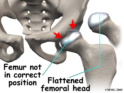 The main problem with Perthes disease is that it changes the structure of the hip joint. How much it affects the way the hip joint works depends on how much the hip joint is deformed. Muscle weakness and atrophy affecting the thigh and calf muscles may develop over time. The affected leg can shorten as a result of the changes in the hip. The result may be a significant leg length difference. Problems later in life are more likely the greater the deformity after the condition has healed.
In general, the most common problem later in life is the development of arthritis in the hip joint. The type of arthritis that develops in the hip is osteoarthritis (also known as wear and tear arthritis). Just like a machine that is out of balance, the hip joint wears out and becomes painful.
The main problem with Perthes disease is that it changes the structure of the hip joint. How much it affects the way the hip joint works depends on how much the hip joint is deformed. Muscle weakness and atrophy affecting the thigh and calf muscles may develop over time. The affected leg can shorten as a result of the changes in the hip. The result may be a significant leg length difference. Problems later in life are more likely the greater the deformity after the condition has healed.
In general, the most common problem later in life is the development of arthritis in the hip joint. The type of arthritis that develops in the hip is osteoarthritis (also known as wear and tear arthritis). Just like a machine that is out of balance, the hip joint wears out and becomes painful.
 Anti-inflammatory medications may also be prescribed. In addition, antiresorptive agents may also be prescribed. These medications help to slow or block the resorption of bone and help decrease deformity. Studies are being done to fully test the effect of these medications in children with Perthes.
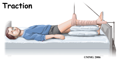
 Physiotherapy while in the hospital is used to restore the hip motion as the inflammation comes under control. A physiotherapist will visit your child in their room and assist them with some gentle hip rotation and abduction exercises (taking the leg out to the side.) These exercises will maintain and improve range of motion but will also assist in moving the fluid inside the hip joint, which assists with joint nutrition and is crucial to healing. They will also show you and your child how to continue the exercises independently once your child leaves the hospital if you will be using a home traction unit, and may prescribe further simple exercises that your child should do once they are no longer in traction. Your physiotherapist may even recommend that your child do some exercises in the pool to take advantage of the hydrostatic properties of the water to gain range of motion with less weight bearing impact.
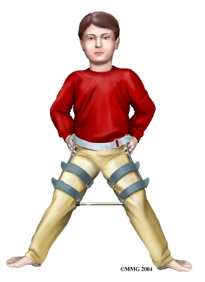
In the past, surgeons have tried to hold the hip in the best position where the femoral head was molded by the acetabulum using many different casts and braces. The most common way of doing this today is the Scottish Rite Orthosis. This brace fits around the waist and thighs and has hinges at the hip joints. The brace allows the child to walk and play while it holds the hip joint in the best position for containment. Your doctor may prescribe this for your child once they leave the hospital. Your physiotherapist will help your child learn to safely use crutches or a walker/frame if they are needed while in the brace.
Physiotherapy while in the hospital is used to restore the hip motion as the inflammation comes under control. A physiotherapist will visit your child in their room and assist them with some gentle hip rotation and abduction exercises (taking the leg out to the side.) These exercises will maintain and improve range of motion but will also assist in moving the fluid inside the hip joint, which assists with joint nutrition and is crucial to healing. They will also show you and your child how to continue the exercises independently once your child leaves the hospital if you will be using a home traction unit, and may prescribe further simple exercises that your child should do once they are no longer in traction. Your physiotherapist may even recommend that your child do some exercises in the pool to take advantage of the hydrostatic properties of the water to gain range of motion with less weight bearing impact.
In the past, surgeons have tried to hold the hip in the best position where the femoral head was molded by the acetabulum using many different casts and braces. The most common way of doing this today is the Scottish Rite Orthosis. This brace fits around the waist and thighs and has hinges at the hip joints. The brace allows the child to walk and play while it holds the hip joint in the best position for containment. Your doctor may prescribe this for your child once they leave the hospital. Your physiotherapist will help your child learn to safely use crutches or a walker/frame if they are needed while in the brace.
 Surgical treatment for containment may be best in older children who are not compliant with brace treatment or where the psychological effects of wearing braces may outweigh the benefits. Surgical containment does not require long-term braces or casts. Once the procedure has been performed and the bones have healed, the child can pursue normal activities as tolerated.
Surgical treatment for containment usually consists of procedures that realign the femur (thighbone), the acetabulum (hip socket), or both.
Surgical treatment for containment may be best in older children who are not compliant with brace treatment or where the psychological effects of wearing braces may outweigh the benefits. Surgical containment does not require long-term braces or casts. Once the procedure has been performed and the bones have healed, the child can pursue normal activities as tolerated.
Surgical treatment for containment usually consists of procedures that realign the femur (thighbone), the acetabulum (hip socket), or both.
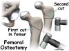
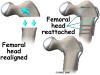
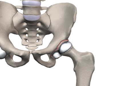
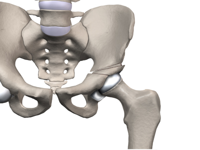
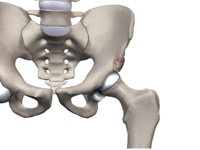
 Realignment of the femur is called a femoral osteotomy. This procedure changes the angle of the femoral neck so that the femoral head points more towards the socket. To perform this procedure, an incision is made in the side of the thigh. The bone of the femur is cut and realigned in a new position. A large metal plate and screws are then inserted to hold the bones in the new position until the bone has healed. The plate and screws may need to be removed once the bone has healed.
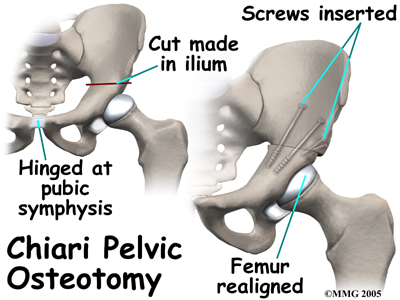
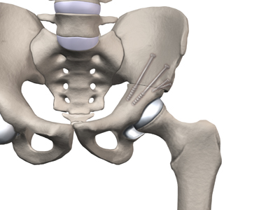
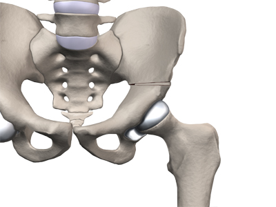
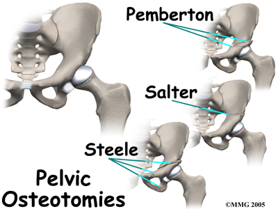
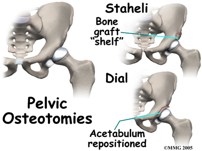 Realignment of the acetabulum is called a pelvic osteotomy. This procedure changes the angle of the acetabulum (socket) so that it better covers, or contains, the femoral head. To perform this procedure, an incision is made in the side of the buttock. The bone of the pelvis is cut and realigned in a new position. Large metal pins or screws are then inserted to hold the bones in the new position until the bone has healed. The pins usually must be removed once the bone has healed.
If there is a serious structural change in the anatomy of the hip, there may need to be further surgery to restore the alignment closer to normal. This is usually not considered until growth stops. As a child grows, there will be some remodeling that occurs in the hip joint. This may improve the situation such that further surgery is unnecessary.
Realignment of the acetabulum is called a pelvic osteotomy. This procedure changes the angle of the acetabulum (socket) so that it better covers, or contains, the femoral head. To perform this procedure, an incision is made in the side of the buttock. The bone of the pelvis is cut and realigned in a new position. Large metal pins or screws are then inserted to hold the bones in the new position until the bone has healed. The pins usually must be removed once the bone has healed.
If there is a serious structural change in the anatomy of the hip, there may need to be further surgery to restore the alignment closer to normal. This is usually not considered until growth stops. As a child grows, there will be some remodeling that occurs in the hip joint. This may improve the situation such that further surgery is unnecessary.
 In severe cases, both femoral osteotomy and pelvic osteotomy may be combined to obtain even more containment.